
Figure 5.
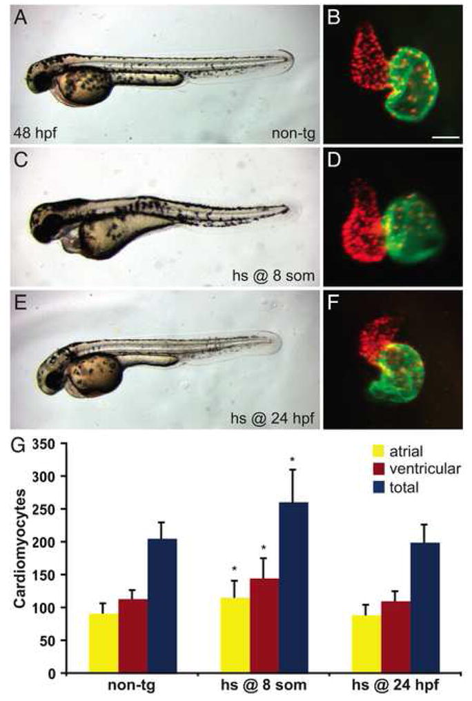
Increased FGF signaling prior to terminal differentiation induces a surplus of cardiomyocytes. (A–F) Representative non-transgenic (A) or Tg(hsp70:ca-fgfr1) (C,E) sibling embryos at 48 hpf, lateral views, exhibiting morphology resulting from heat shock at the 8-somite stage (C) or at 24 hpf (E), coupled with respective frontal views of hearts in which immunofluorescence detects DsRed (red) in all cardiomyocyte nuclei and Amhc (green) in atrial cells, as in Figure 1. Scale bar represents 50 μm; all images of hearts are shown at the same magnification. (C,D) Although heat shock at the 8-somite stage causes pericardial edema (C) and dysmorphic cardiac chambers (D), heat shock at 24 hpf does neither (E,F). (G) Quantification of cardiomyocytes at 48 hpf reveals that increased FGF signaling causes a significant cardiomyocyte surplus only in embryos heat shocked at the 8-somite stage. Graph indicates mean and standard deviation for each data set; asterisks indicate statistically significant differences relative to non-tg controls (p<0.005, Student’s t-test). n=24 for non-tg, n=18 for heat shock at the 8-somite stage, and n=14 for heat shock at 24 hpf; see also Supplemental Table 5.