
Figure 7.
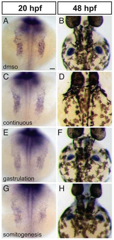
Reduction of FGF signaling does not expand the forelimb field. (A–H) In situ hybridization depicts expression of tbx5 at 20 hpf (A,C,E,G) and 48 hpf (B,D,F,H); dorsal views, anterior to the top. Scale bar represents 50 μm; all images are shown at the same magnification. (A–D) Compared to control embryos treated only with DMSO (A,B), embryos exposed to continuous SU5402 treatment exhibit normal areas of tbx5 expression in the pectoral fin fields at 20 hpf (C; n=8/8) but lack pectoral fins at 48 hpf (D; n=15/15). (E,F) Embryos treated with SU5402 during gastrulation exhibit normal areas of tbx5 expression in the pectoral fin fields at 20 hpf (n=16/16) and normal pectoral fins at 48 hpf (n=18/18). (G,H) Embryos treated with SU5402 during somitogenesis do not display increased areas of tbx5 expression in the pectoral fin fields at 20 hpf (n=9/9) and do not have detectable defects in pectoral fin size at 48 hpf (n=15/16), although fin morphology varies between treated embryos. It is noteworthy that the level of tbx5 expression generally appears higher in wild-type embryos than in SU5402-treated embryos.