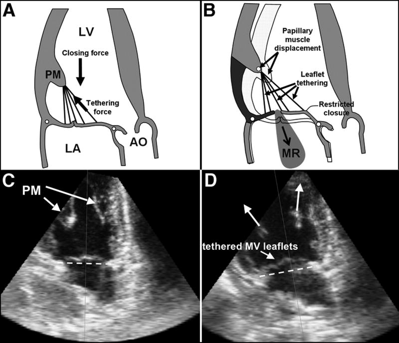
Figure 1.
Normal MV closure (A,C); effect of apical and posterior shift of PMs restraining the MV, causing MV leaflet tethering (D) and, if severe enough, MR (B). Lower panel (C & D): Echocardiography of the LV (apical two-chamber view) in the same sheep prior to (C) and after pulling the PMs apically resulting in MV leaflet tethering (D). The dashed line indicates the mitral annular level. (Ao: Aorta, LA: left atrium; LV: left ventricle, MR: mitral regurgitation; MV: mitral valve; PM: papillary muscle) (Figures A & B adapted from Levine et al 14).

