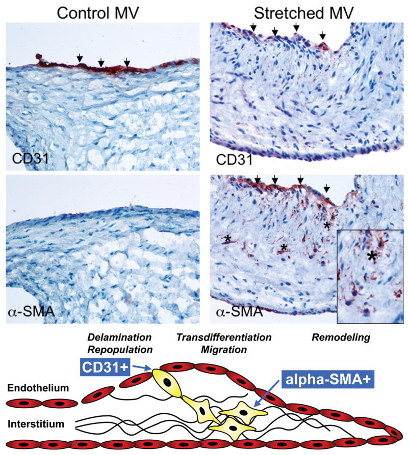
Figure 6.
Left: Unstretched MV showing negative α-SMA staining along the CD31+ endothelium. Right: α-SMA+ staining in the atrial endothelium (also CD31+) of a stretched MV, with nests of α-SMA+ cells appearing to penetrate the interstitium (asterisks, below). Lower panel: Schematic of active mitral valve adaptation by EMT (adapted from Armstrong et al 36). (α-SMA: α-smooth muscle actin)

