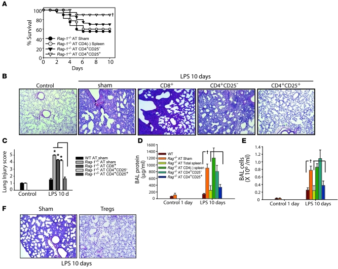
Figure 3. AT of Tregs mediates resolution of lung injury in Rag-1–/– mice.
Rag-1–/– mice were challenged with i.t. LPS and 1 hour afterward received PBS sham treatment or 1.0 × 10⁶ WT CD4+CD25– or WT CD4+CD25+ splenocytes. (A) Rag-1–/– mouse survival over a 10-day period. †P < 0.05 versus sham control, log-rank test. (B) H&E stain of representative lung sections on day 10 after i.t. LPS and infusion of PBS or the indicated lymphocyte subsets. Original magnification, ×40. (C) Mean histopathological lung injury scores (n = 8–10 animals per group). *P < 0.05. (D and E) BAL total protein (D) and total cell counts (E) were determined in WT and Rag-1–/– mice on day 10 after AT (n = 10 per group). †P < 0.05 versus Rag-1–/– sham control. (F) Lung H&E staining demonstrate that AT of Tregs into injured Rag-1–/– mice as late as 24 hours after i.t. LPS achieved resolution of lung injury (n = 5 per group). Original magnification, ×40.

