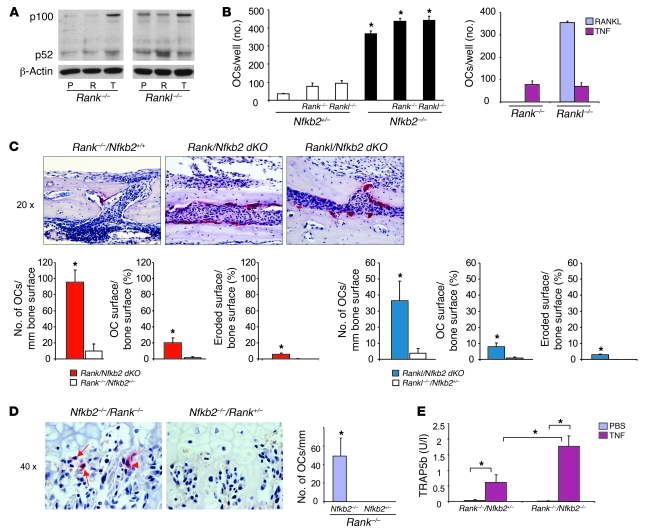
Figure 2. NF-κB2 deficiency enhances TNF-induced osteoclastogenesis in Rank–/– or Rankl–/– mice in vitro and in vivo.
(A) NF-κB p100 and p52 were analyzed by Western blot in whole-cell lysates of PBS-, RANKL-, or TNF-treated (8 hours) OCPs from Rank–/– or Rankl–/– mice. (B) Left: OCPs from Rank–/–/Nfkb2–/– or Rankl–/–/Nfkb2–/– mice and their Nfkb2+/– littermates were treated with TNF for 2 days to evaluate osteoclast formation using TRAP staining (*P < 0.05 vs. Nfkb2+/–). Right: OCPs from Rank–/–/Nfkb2+/+ and Rankl–/–/Nfkb2+/+ mice were treated with RANKL or TNF for comparison with Rank–/–/Nfkb2+/– and Rankl–/–/Nfkb2+/– mice to determine the effects of haploinsufficiency of Nfkb2. (C) Murine TNF (0.5 μg in 10 μl PBS) or 10 μl PBS was injected twice daily over the calvariae of Rank–/–/Nfkb2–/– or Rankl–/–/Nfkb2–/– mice and Rank–/– or Rankl–/– littermates. Top: TRAP-stained sections show numerous actively resorbing TRAP+ osteoclasts locally in calvarial sections (original magnification, ×20) from TNF-injected Rank–/–/Nfkb2–/– or Rankl–/–/Nfkb2–/– mice. Bottom: Numbers and surface extent of osteoclasts (n = 3/genotype). Occasional osteoclasts induced by TNF from a Rank–/–/Nfkb2+/+ mouse are illustrated in the left panels. *P < 0.05 vs. single KO mice. (D) Left: Occasional binucleate (arrowhead), but mainly mononuclear (arrows), TRAP+ cells (left panel) formed beneath hypertrophic chondrocytes in the growth plate of the tibia of a Rank–/–/Nfkb2–/– mouse (original magnification, ×40), but not of Rank–/–/Nfkb2+/– littermates injected with TNF as described in C. Right: Osteoclast numbers (expressed per mm of length of growth plate) counted in representative sections. (E) Serum TRAP5b levels were tested with ELISA from TNF- or PBS-injected Rank–/–/Nfkb2–/– and Rank–/–/Nfkb2+/– mice (n = 3/group; *P < 0.05).

