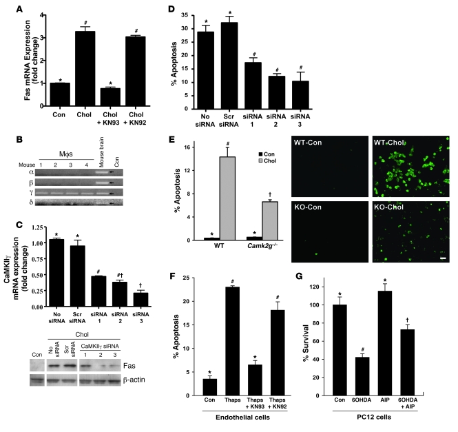
Figure 3. Induction of Fas and apoptosis by ER stress involves CaMKII.
(A) Macrophages were incubated for 8 hours under cholesterol-loading conditions with or without KN93 or KN92 after 1 hour pretreatment, and then assayed for Fas mRNA. (B) RNA from peritoneal macrophages from 4 separate mice and from mouse brain, along with water control, were probed for the indicated CaMKII isoform mRNAs by RT-PCR. (C) Macrophages were transfected with 3 different CaMKIIγ siRNA constructs. After 72 hours, the cells were incubated for 8 hours under cholesterol-loading conditions and then assayed for CaMKIIγ mRNA and total Fas protein. (D) Macrophages were transfected with the 3 siRNA constructs in C, incubated for 30 hours with 0.25 μM thapsigargin and 25 μg/ml of the SRA ligand fucoidan, and then assayed for apoptosis. (E) Macrophages from WT or Camk2g–/– mice were incubated under control or cholesterol-loading conditions for 12 hours and then assayed for apoptosis. Scale bar: 20 μm. (F) Human aortic endothelial cells were incubated for 24 hours with thapsigargin (1 μM) with or without KN93 or KN92 after 1 hour pretreatment, and then assayed for apoptosis. (G) PC12 cells were incubated for 24 hours with 100 μM 6-OHDA with or without 10 μM AIP-II after 1 hour pretreatment, and then assayed for cell viability (percentage of viable cells compared with those in cultures not treated with 6-OHDA). Differing symbols indicate P < 0.01; identical symbols indicate differences that are not significant.

