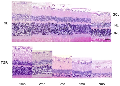
Figure 1. Illustration of histological assessment for neuronal changes in TGR rats from 1 to 7 months.
Representative images showing progressive degenerating photoreceptor cells in the TGR rats compared to age-matched SD rats. Note that the SD rats have obvious photoreceptor cell loss during ageing. n = 3−8. Upper: SD rats; lower: TGR rats. GCL: ganglion cell layer; INL: inner nuclear layer; ONL: outer nuclear layer. Scales: 25 µm.

