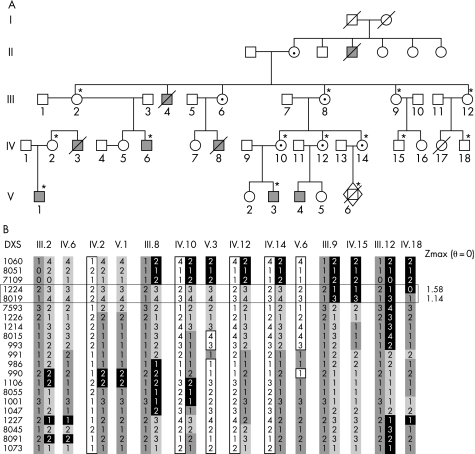
Figure 1 (A) Pedigree of the family included in the linkage analysis. *Members for whom DNA was available and who were included in the linkage analysis. Filled squares, affected male patients; circles with dots, heterozygous female members. Female carrier status was assigned from family history (obligate carriers). (B) Haplotypes of the X‐chromosome markers and the recombined markers delimiting the potential genetic interval (horizontal rectangle). Maximum LOD scores are indicated on the right.

An official website of the United States government
Here's how you know
Official websites use .gov
A
.gov website belongs to an official
government organization in the United States.
Secure .gov websites use HTTPS
A lock (
) or https:// means you've safely
connected to the .gov website. Share sensitive
information only on official, secure websites.
