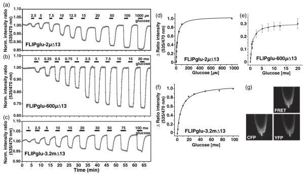
Figure 1.
Glucose-induced FRET changes in the cytosol of intact roots.
(a-c) The FRET sensors FLIPglu-2μΔ13 (a), FLIPglu-600μΔ13 (b), and FLIPglu-3.2mΔ13 (c) respond to glucose perfusion in stably transformed rdr6-11 Arabidopsis roots after 5, 3 or 2 days of transfer to sugar-free medium, respectively. Quantitative data were derived by pixel-by-pixel integration of the ratiometric images. The y axis gives the ratio of eYFP intensity (ET535/30m) to eCFP intensity (ET470/24m). The bars above each graph represent the duration of perfusion with the indicated sugar concentrations.
(d-f) Saturation curves derived from the data shown in (a)-(c) for FLIPglu-2μΔ13 (d), FLIPglu-600μΔ13 (e) and FLIPglu-3.2mΔ13 (f).
(g) Confocal images showing the expression pattern of FLIPglu-600μΔ13 in the CFP, YFP and FRET channels in root tips.

