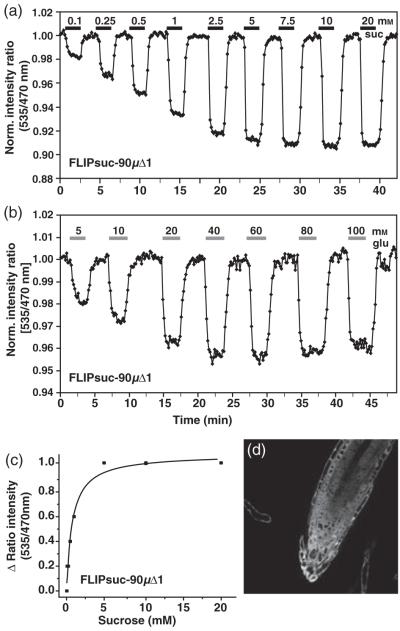
Figure 5.
Sucrose-induced FRET changes in the cytosol of intact roots.
(a.b) The FRET sensor FLIPsuc-90μΔ1 responds to sucrose perfusion (a) and glucose perfusion (b) in stably transformed rdr6-11 Arabidopsis roots. Images were acquired and data analyzed as in Figure 1. The bars above each graph represent the duration of perfusion with the indicated sucrose concentrations (black) or glucose concentrations (grey).
(c) Saturation curve for a representative in vivo sucrose titration of FLIPsuc-90μΔ1.
(d) Expression pattern (YFP channel) of FLIPsuc-90μΔ1 in root tips.

