
Fig. 4.
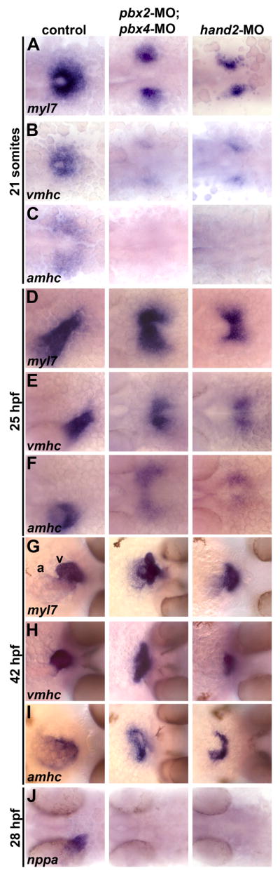
Pbx-deficient and hand2-MO embryos exhibit similar cardiac differentiation and morphogenesis defects. (A–J) RNA in situ expression of (A,D,G) myl7, (B,E,H) vmhc, (C,F,I) amhc, and (J) nppa at (A–C) 21 somites, (D–F) 25 hours post fertilization (hpf), (G–I) 42 hpf, and (J) 28 hpf. In control embryos shown, myl7 labels all myocardial precursors, vmhc labels ventricular precursors, and amhc labels atrial precursors (Berdougo et al., 2003). Ventricle (v) and atrium (a) are labeled in (G). n≥20 for each marker in control, pbx2-MO; pbx4-MO, or hand2-MO. (A–F) Embryos are shown in dorsal view, anterior towards the left. (G-I) Embryos are shown in frontal views, dorsal towards the right.