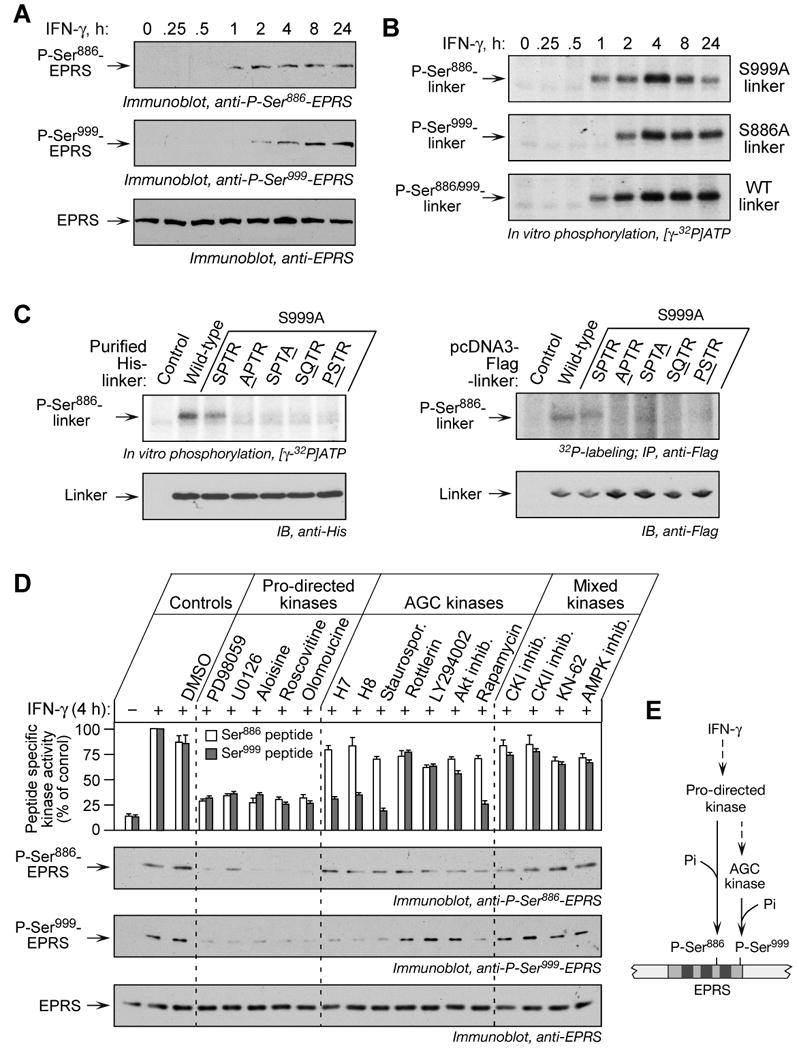
Figure 3. Ordered Phosphorylation of Ser886 and Ser999 by two Ser/Thr kinases.

(A) Ser886 phosphorylation precedes Ser999 phosphorylation. Lysates from U937 cells treated with IFN-γ were resolved by SDS-PAGE (12% polyacrylamide) and analyzed by immunoblot with phospho-specific antibodies, and with anti-EPRS antibody.
(B) Ser886 and Ser999 phosphorylation events are independent. Lysates of IFN-γ-treated U937 cells were used to in vitro phosphorylate His-tagged wild-type and mutant linkers.
(C) Identification of the kinase recognition motif for Ser886 phosphorylation. Lysates of 4-h, IFN-γ-treated cells were used to in vitro phosphorylate purified His-tagged wild-type and linkers with mutations in key motif amino acids; equal linker amount was detected with anti-His antibody (left panels). The pcDNA3-Flag-linker DNAs containing motif mutations were transiently expressed in U937 cells and labeled with 32P-orthophosphate in presence of IFN-γ for 4 h. Lysates were immunoprecipitated with anti-Flag antibody, and 32P incorporation determined by autoradiography (right panels).
(D) Distinct kinases phosphorylate Ser886 and Ser999. U937 cells were incubated with IFN-γ for 0.5 h and then with kinase inhibitors for an additional 3.5 h. Cells incubated with or without IFN-γ and dimethyl sulfoxide (DMSO, 0.5%) served as controls. Pro-directed kinase group inhibitors were PD98059 (10 μM), U0126 (10 μM), aloisine (5 μM), roscovitine (10 μM), and olomoucine (50 μM). AGC kinase group inhibitors were H-7 (10 μM), H-8 (10 μM), staurosporine (Staurospor. 20 nM), rottlerin (100 μM), LY294002 (2.5 μM), Akt inhibitor-VIII (2.5 μM), rapamycin (10 nM). Other inhibitors were CKI inhibitor (100 μM), CKII inhibitor (100 μM), KN-62 (5 μM), and AMPK inhibitor (2 μM). Lysate kinase activity was determined as phosphorylation of Ser886 (white) and Ser999 (grey) phospho-acceptor peptides, and expressed as % of control lysates (mean ± SEM, top panel). Lysates were resolved on SDS-PAGE (12% polyacrylamide) and analyzed by immunoblot with indicated antibodies (panels 2-4).
(E) Schematic of kinase pathway phosphorylating EPRS Ser886 and Ser999.
