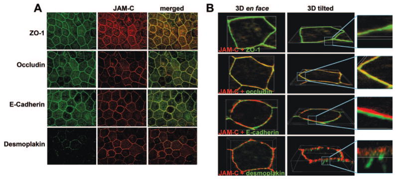
Figure 2.
Localization of JAM-C in the cultured hfRPE cells. hfRPE monolayers were double stained with anti-JAM-C (red) and with anti-ZO-1, anti-occludin, anti-E-cadherin, and anti-desmoplakin (green). (A) Junctional localization of JAM-C. (B) En face and tilted views of 3-D reconstruction of single cells, showing that JAM-C colocalized with ZO-1 and occludin and was situated apically to E-cadherin and desmoplakin. Similar results were obtained in three experiments.

