
Figure 5.
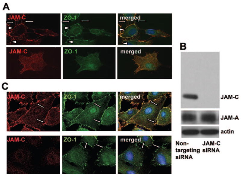
Role of JAM-C in the recruitment of ZO-1 to the initial hfRPE contacts. (A) Double staining for JAM-C (red) and ZO-1 (green) in contacting hfRPE cells show recruitment of JAM-C at the contacting surfaces of adjacent cells, similar to the distribution of ZO-1. In noncontacting hfRPE cells, immunofluorescence for both JAM-C and ZO-1 showed no specific staining. Similar results were obtained in three experiments. (B) Western blot analysis of hfRPE cells transfected with siRNA against JAM-C or with control siRNA as indicated, show the expression of JAM-C, JAM-A, and actin. JAM-A expression was not changed after JAM-C knockdown. (C) JAM-C knockdown caused a disruption in the ZO-1 localization at the initial contacts among hfRPE cells (bottom) compared with the control transfected cells (top). Similar results were obtained in three experiments.