
Figure 6.
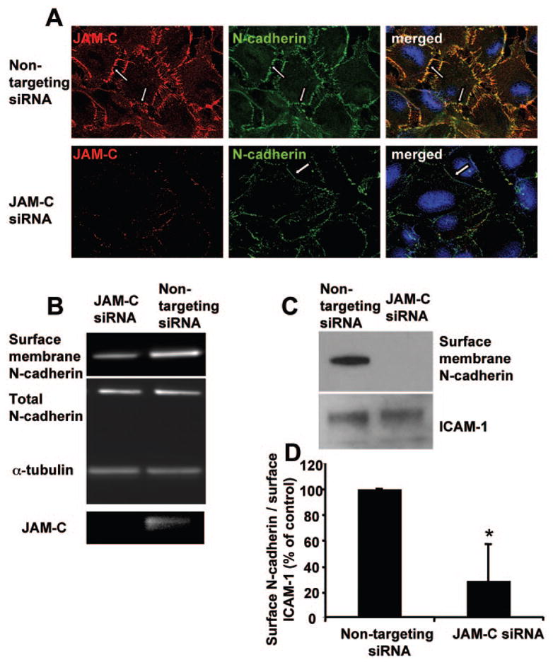
Effect of JAM-C in the recruitment of N-cadherin at the initial hfRPE contacts. (A) JAM-C and N-cadherin staining of control siRNA transfected and JAM-C siRNA transfected hfRPE cells. (B) Western blot analysis for the surface membrane fraction of N-cadherin, total N-cadherin, α -tubulin, and JAM-C in hfRPE cells transfected with control nontargeting siRNA or specific JAM-C targeting siRNA. A reduction in surface-associated N-cadherin but no change in total N-cadherin was observed on JAM-C knockdown. (C) Western blot analysis for the surface membrane fraction of N-cadherin and the surface membrane fraction of ICAM-1 in hfRPE cells transfected with control nontargeting siRNA or specific JAM-C targeting siRNA. (D) Densitometric analysis of the surface membrane fraction of N-cadherin and the surface membrane fraction of ICAM-1 in hfRPE cells transfected with control nontargeting siRNA or specific JAM-C targeting siRNA. Data are shown as a percentage of control and represent the ratio of surface membrane fraction of N-cadherin to the surface membrane fraction of ICAM-1 in cells transfected with nontargeting siRNA (mean ± SD; n = 3; *P < 0.05).