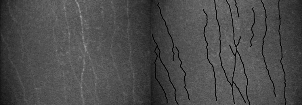
Figure 1.

Subbasal nerve density measurement. (Left) Subbasal nerves in a normal cornea as recorded by using Tandem Scanning confocal microscope and (right) the same image after tracing nerves by using a semi-automated nerve analysis program. 8 Subbasal nerve density (µm/mm2) was the total length of nerve (µm) measured per image sample area (0.058 mm2 for the ConfoScan 4 and 0.166 mm2 for Tandem Scanning).
