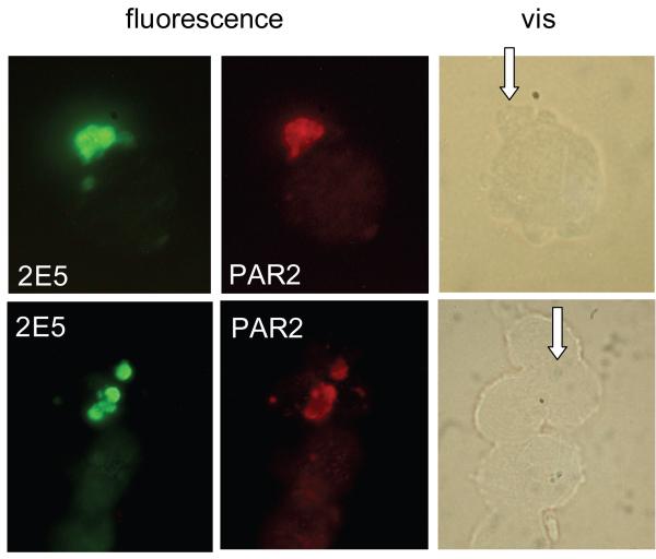
Fig. 2. Immunofluorescence analysis of PAR2 expression on infected cells.
Cells recovered from a HCT-8 monolayer 24 h post-infection were double-labelled for C. parvum (2E5 monoclonal antidoby) and PAR2 as indicated. In unlabelled cells the parasites are difficult to discern and their position are indicated with arrows.

