Figure 3.

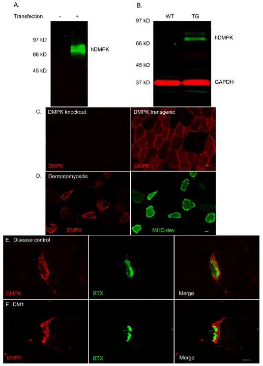
DMPK protein localization in muscle tissue. (a) Antibody 5244 recognized hDMPK by immunoblot of COS-7 cells transfected with hDMPK expression construct. (b) This antibody also recognized hDMPK by immunoblot in lysates prepared from hDMPK transgenic mouse muscle, but not in wild type muscle. (c) Immunofluorescence reveals cytoplasmic hDMPK in muscle sections from hDMPK transgenic mouse that is absent in DMPK knockout mouse muscle. (d) DMPK (red) is upregulated in regenerating fibers labeled with developmental myosin (green), shown here in dermatomyositis. (e,f) DMPK protein (red) accumulates at the NMJ in DM1 and disease control muscle, localizing to a subsynaptic domain immediately beneath the acetylcholine receptors labeled by α-BTX (green). Bar = 10 μM.
