
Figure 1.
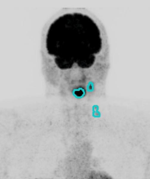
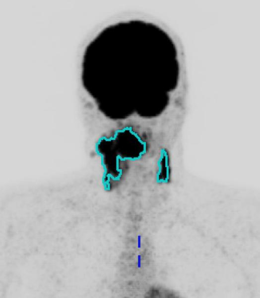
Primary site and regional nodal metabolic tumor volume. Maximum intensity projection views of 18F-fluorodeoxyglucose-positron emission tomography scans for two patients, with overlay of segmented metabolic tumor volumes (MTV). Figure 1a. Patient with Stage IVA oropharyngeal cancer (T3N2bM0) with small MTV (MTV: 63.3 ml, Progression free survival: 30.9 months). Figure 1b. A patient with Stage IVB (T4bN2cM0) oropharyngeal cancer with large MTV (MTV: 511.6 ml, Progression free survival: 13.8 months).