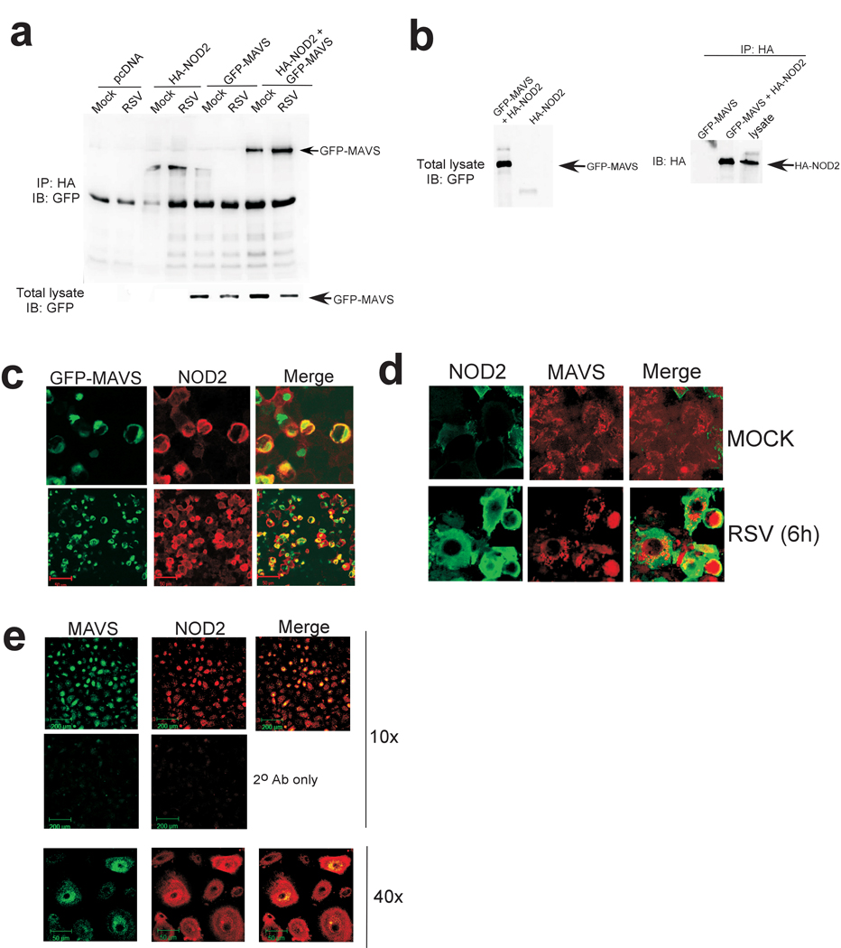
Figure 6.

Interaction of MAVS with NOD2. (a) 293 cells were transfected with pcDNA, HA-NOD2 and-or GFP-MAVS and mock infected or infected with RSV. Lysates were immunoprecipitated with anti-HA agarose beads and bound proteins were immunoblotted with anti-GFP. Expression of GFP-IPS-1 in the cell lysate (by immunoblotting 25 µg of total cellular lysate with anti-GFP antibody) is also shown in the lower panel. (b) Expression of GFP-IPS-1 and HA-NOD2 in the cell lysate (by immunoblotting with anti-GFP and anti-HA antibodies) and amount of HA-NOD2 bound to anti-HA-agarose beads is also shown. For immunoblotting with cell lysates, 25 µg of total cellular lysate protein was used to detect GFP-IPS-1 and HA-NOD2. (c) RSV-infected (4h) 293 cells co-expressing GFP-MAVS (green) and HA-NOD2 (red) were imaged using confocal microscopy. (d,e) Mock or RSV-infected (6h) A549 cells (d) or RSV-infected (4h) NHBE cells (e) were stained with anti-NOD2 and anti-MAVS and imaged by confocal microscopy to detect endogenous NOD2 and MAVS.
