
Figure 4.
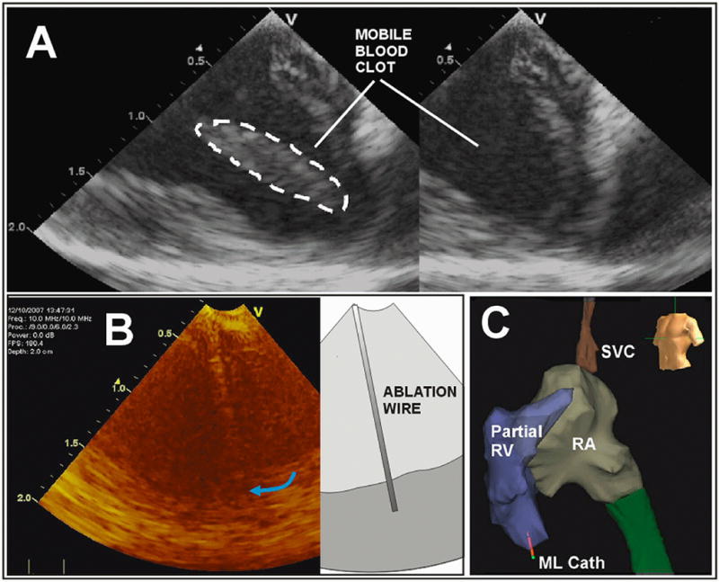
The ML in-vivo imaging image quality is demonstrated in A with a display depth of 2cm. A special ablation wire is tracked in B, and the ML catheter tip 3D spatial position in the apical region of the RV is tracked dynamically in panel C with a partially completed volumetric map of the right side of the pig heart with the use of the NavX electroanatomical mapping system; note the ML catheter tip shown near the RV apex at the bottom left in C.