
Figure 7.
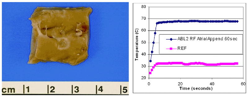
An ablation lesion created on the endocardial surface of the right atrial appendage is shown at the left, with the ablation site temperature plot at right. To confirm tissue temperatures during ablation a pair of 0.5mm fluoroptic (Luxtron, Santa Clara, Calif.) temperature probes were placed within the myocardium.