Fig. 2.
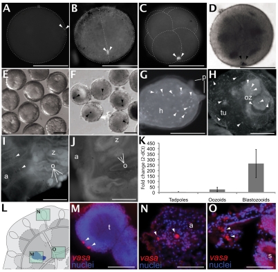
vasa expression during embryogenesis, metamorphosis and in adult colonies as shown by ISH. (A) Fertilized egg with polar bodies in the animal pole and granule-like aggregation of maternal vasa (arrowheads) in the cortex. (B) A two-cell-stage embryo with vasa mRNA (arrowheads) aggregated on one side of the cleavage furrow. (C) An eight-cell-stage embryo with two proximate aggregates of vasa (arrowheads) at the posterior cortical region of the B4.1 blastomeres. (D) Sixteen-cell-stage embryo with vasa mRNAs (arrowheads) at the posteriormost cortical region of the B5.1 blastomeres. (E) Gastrula- and neural-plate-stage embryos (past the 110-cell stage) show invagination and blastopore formation on the vegetal side of the embryo. (F) Gastrula- and neural-plate-stage embryos with a single aggregate of vasa (arrowheads) in the posterior region of the embryos. (G) Detail of a larval head during metamorphosis. Anterior adhesive papillae (p) can be observed on the right; vasa+ cells (arrowheads) are scattered throughout the head (h). (H) vasa expression in a newly settled colony is seen in individual cells (arrowheads) in the oozooid (oz), and within the extracorporeal vasculature. (I) In a sexually mature adult, vasa expression is seen in small oocytes (o) at the site of the gonads of the zooid (z) and in individual cells (arrowheads) within the ampullae (a). (J) A sexually fertile adult shows general background levels of autofluorescence in blood and tunic cells, as shown by the sense probe negative control (cf. H and I; antisense probe). (K) Quantitative PCR analysis of vasa mRNA levels during metamorphosis. Larvae show the lowest mRNA levels, oozooids show a slight increase, and first-generation asexually derived zooids show a further increase after ten days of settlement. (L) Illustration of a colony (dorsal view) showing location of the testis, ampullae and zooid body (framed), corresponding to histological sections in M-O. (M) Fluorescence in situ hybridization shows two vasa expressing cells (red; arrowheads) at the periphery of a mature testis containing highly packed nuclei of sperm precursors (blue). (N) vasa expressing cells (red, arrowheads) in the ampullae. vasa+ cells are scattered throughout the vasculature and are surrounded by other hemocytes (blue, nuclei counterstained with DAPI). (O) Cells expressing vasa (red, arrowheads) are also seen in the sinuses and lacunae surrounding the epithelial folds of the stomach (s) in an adult zooid of the colony. a, ampullae; h, head; o, oocyte; oz, oozoid; p, papillae; s, stomach; t, testis; tu, tunic; z, zooid. Scale bars: 100 μm.

