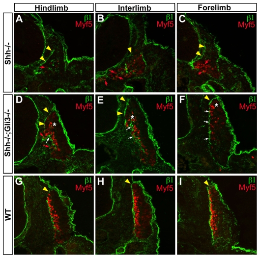
Fig. 5.
Progressive recovery of myotomal basement membrane in Gli3-/-;Shh-/- embryos. (A-I) β1 laminin subunit (green) and Myf5 (red) protein distribution examined by immunohistochemistry and confocal imaging in somites of E10.0 Shh-/- (A-C), Shh-/-;Gli3-/- (D-F), and wild-type (G-I) embryos. (A-C) Shh-/- embryos. (D-F) Shh-/-;Gli3-/- somites. Note the recovery of epaxial Myf5 activation at all axial levels (white asterisk). (G-I) Wild-type embryos. Yellow arrowheads indicate the dermomyotomal basement membrane. Red arrows show abnormally located myotomal cells. White arrows indicate sites of partially assembled basement membrane. Magnification: ×400.

