Fig. 7.
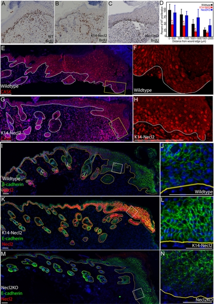
Necl2 overexpression decreases proliferation and leads to upregulation of E-cadherin and CASK during wound healing. (A-C) BrdU-labelled S-phase cells (brown, A-C) at the leading edge of healing wild-type (A), transgenic (B) and Necl2-knockout (C) mouse epidermis. (D) Quantification of BrdU-positive cells as a function of distance from the wound edge in wild-type (black bars), transgenic (red bars), and knockout mice (blue bars). Data are presented relative to appropriate congenic wild-type controls (n=4 knockout mice/group; n=8 transgenic mice/group). Error bars show s.e.m.; *P<0.05. (E-H) Wounded wild-type (E,F) or K14Necl2 transgenic (G,H) epidermis stained with anti-CASK (red) and DAPI nuclear counterstain (blue; E,G). (I-N) E-cadherin (green, I-N) and Necl2 (red, I,K,M) expression at the leading edge of re-epithelialising wounds, 7 days after wounding. (E,G,I-N) DAPI nuclear counterstain (blue). Lines (A-C,E-N) denote dermal-epidermal boundary and boxes (E,G,I,K,M) denote regions shown at higher magnification in adjacent panels. Scale bars: 50 μm in A-C; 100 μm in E-N.

