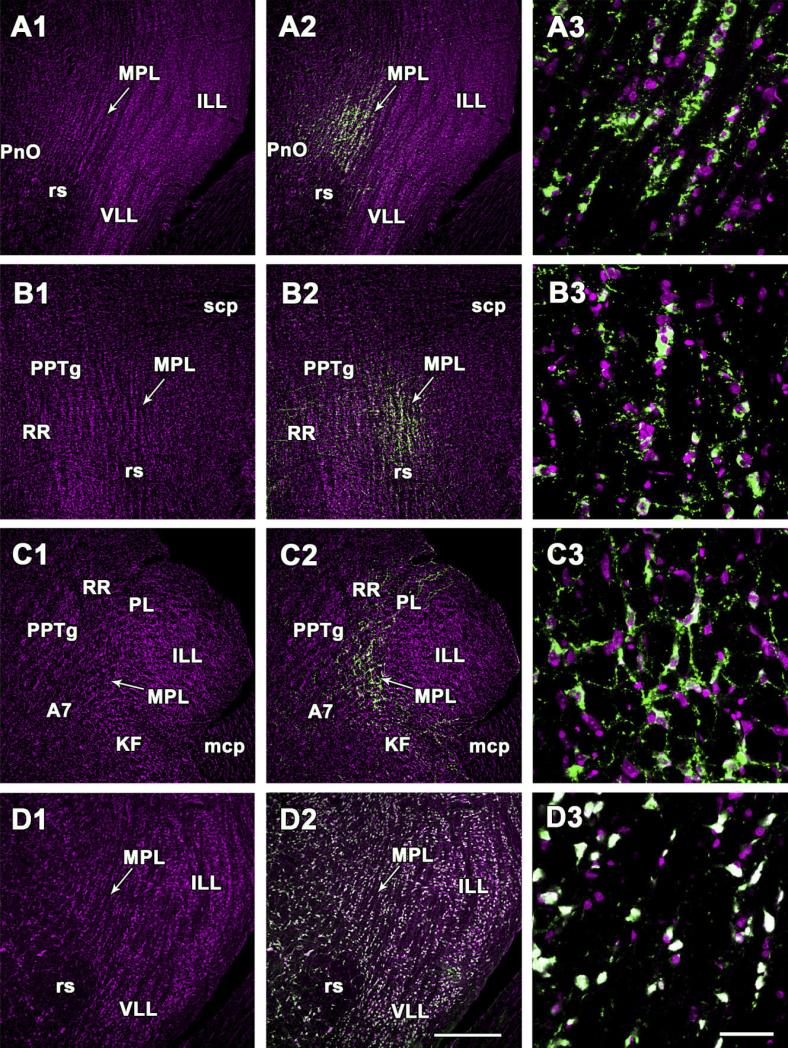
Fig. 2.
Demonstration of TIP39- and NeuN-immunoreactive neurons in the cytoarchitectonically defined medial paralemniscal nucleus. A1–A3, D1–D3: Coronal sections. B1–B3: Sagittal sections. C1–C3: Horizontal sections. A1,B1,C1,D1: Sections are stained with the red fluorescent Nissl dye, and the picture is artificially made magenta. Arrows point to the medial paralemniscal nucleus. A2,B2,C2: The position of TIP39 neurons is demonstrated by green fluorescent immunolabeling in the same field of the same sections as A1, B1, and C1. D2: The same field as D1 is shown double labeled with the red fluorescent Nissl dye (magenta) and NeuN (green). The Nissl dye and NeuN co-localizes in all the neurons indicated by their white color. A3, B3, C3, and D3 are high-magnification confocal photomicrographs. The fields correspond to MPL in the middle panels. For abbreviations, see list. Scale bar = 500 μm in D2 (applies to A1–D2); 100 μm in D3 (applies to A3–D3).

