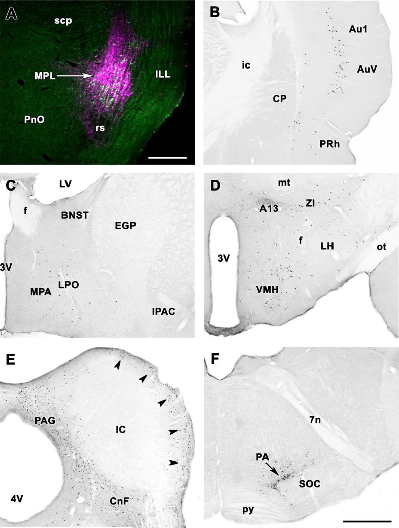
Fig. 4.
Topographical distrbution of retrogradely labeled neurons after CTB injection into the MPL. A: The CTB injection site (magenta) is shown in relation to TIP39-immunoreactive neurons (green) in a double fluorescent labeled coronal section. The injection site covers most of the MPL. B–F: Photomicrographs of coronal sections single labeled with DAB immunocytochemistry demonstrate retrogradely labeled neurons in the same brain. B: A high density of retrogradely labeled neurons is shown in layer V of the primary auditory cortex and the ventral secondary auditory cortex. Labeled cells in a lower density are present in layers Vi and V of the perirhinal cortex and layer VI of the ventral secondary auditory cortex. C: Retrogradely labeled neurons are distributed in the preoptic region. D: A high density of retrogradely labeled neurons in the ventrolateral subdivision of the hypothalamic ventromedial nucleus, the dorsolateral hypothalamic area, the area of the A13 dopamine cell group, and the zona incerta. Scattered cells are also present in the lateral hypothalamus. E: Retrogradely labeled neurons are abundant in the dorsal periaqueductal gray, the cuneiform nucleus, and the external cortex of the inferior colliculus (arrowheads). In contrast, the central nucleus of the inferior colliculus is devoid of labeled neurons. F: Retrogradely labeled cells are concentrated in the periolivary area immediately dorsal to the superior olive. For abbreviations, see list. Scale bar = 500 μm in A; 1 mm in F (applies to B–F).

