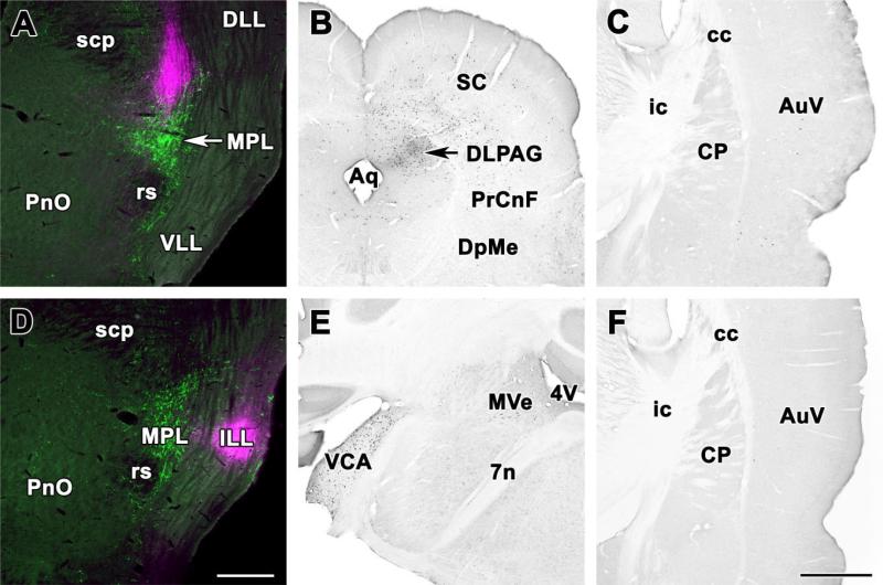
Fig. 6.
Location of retrogradely labeled neurons following control injections into areas adjacent to the MPL. A: Injection into the paralemniscal region immediately dorsal to the MPL. The CTB injection site (magenta) overlaps only slightly with the MPL containing TIP39-immunoreactive neurons (green). B: Photomicrographs of coronal sections single labeled with DAB immunocytochemistry demonstrate retrogradely labeled neurons in the periaqueductal gray and the superior colliculus of the same brain. C: In contrast, the auditory cortex contains only a few labeled neurons. D: Injection into the intermediate nucleus of the lemniscus lateralis. The CTB injections site (magenta) is located lateral to the MPL containing TIP39-immunoreactive neurons (green). E: Photomicrographs of coronal sections single labeled with DAB immunocytochemistry demonstrate a very high number of retrogradely labeled neurons in the contralateral ventral cochlear nucleus and a less moderate labeling in the magnocellular medial vestibular nucleus of the same brain. F: In contrast, the auditory cortex contains no labeled neurons. For abbreviations, see list. Scale bar = 500 μm in D (applies to A,D); 1 mm in F (applies to B,C,E,F).

