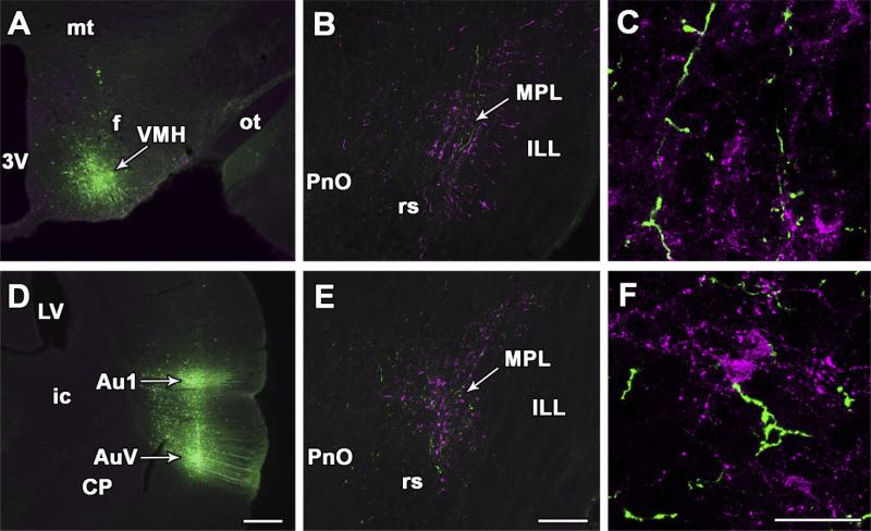
Fig. 7.
Projections from the hypothalamic ventromedial nucleus and the auditory cortex to the MPL. A: BDA injection site in the ventrolateral subdivision of the hypothalamic ventromedial nucleus. B: In the same brain, anterogradely labeled fibers (green) are distributed among TIP39-immunoreactive neurons (magenta) in the MPL. C: High-magnification confocal images suggest BDA-containing nerve terminals in the MPL. D: BDA injection sites in the primary and secondary auditory cortices. E: In the same brain, anterogradely labeled fibers (green) are distributed among TIP39-immunoreactive neurons (magenta) in the MPL. F: High-magnification confocal images suggest BDA-containing nerve terminals in the MPL. For abbreviations, see list. Scale bar = 500 μm in D (applies to A,D); 300 μm in E (applies to B,E); 50 μ in F (applies to C,F).

