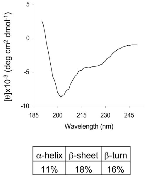
Figure 6. Circular dichroism (CD) illustrates secondary structural features of proenkephalin (PE).
Analysis of the CD spectrum of PE revealed secondary structures of PE. The CD spectrum of recombinant PE was obtained, and was deconvoluted using CDSSTR (21) to reveal secondary structures consisting of 11% alpha-helix, 18% beta-sheet, 16% beta-turn, and 55% disordered structures.

