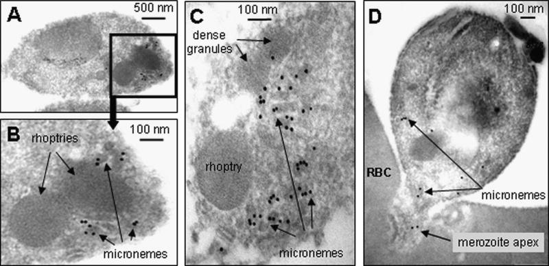
Figure 3.
Immuno-electron microscopic localization of the PkRBL protein, PkNBPXb. In A – C, PkNBPXb is shown to be strongly localized to the apical micronemes of free P. knowlesi merozoites, using rabbit anti- NBPXb anti-serum (1:50) as primary antibody, and Protein A -10 nm gold for detection.. In an invading merozoite (D), very little labelling remains (rabbit anti-NBPXb anti-serum 1:100 dilution used), and is mostly detected on micronemes that have apparently failed to locate apically, suggesting that the RBL had already been mostly secreted and lost, as expected if the protein is important in red cell adhesion. Note the absence of general labelling around the merozoite perimeters in all examples. RBC – red blood cell.

