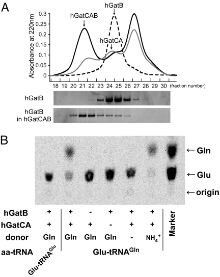
Fig. 3.
In vitro reconstitution of Gln-tRNAGln formation by hGatCAB. (A) Gel filtration chromatography (Superdex 200) of hGatCA (gray line), hGatB (dotted line), and a mixture of hGatCA and hGatB (black line) detected by UV absorption at 220 nm. Elution profiles of hGatB alone (Upper) and hGatB in the hGatCAB complex (Lower) were analyzed by Western blotting using an anti-His-tag antibody. (B) In vitro reconstitution of Gln-tRNAGln formation by the recombinant hGatCAB. Phosphor-image of the TLC analysis of [14C]-labeled Gln and Glu deacylated from aa-tRNAs in transamidation experiments. The assays were performed in the presence (+) or absence (−) of hGatB or hGatCA. Gln or NH4+ was used as an amide donor. [14C]-labeled Glu-tRNAGlu or Glu-tRNAGln was used as a substrate, as indicated below the panel. Positions of Gln and Glu on TLC were determined using [14C]Gln and [14C]Glu as markers (right lane).

