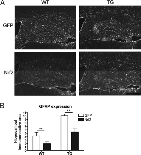
Fig. 5.
APP/PS1 mice exhibit elevated astrogliosis that is attenuated by LV-Nrf2 treatment. (A) Representative photomicrographs of astrocytic GFAP immunostaining in hippocampus of mice injected with LV-GFP or LV-Nrf2. (Scale bar, 200 μm.) (B) APP/PS1 mice exhibit elevated amounts of GFAP immunoreactivity. Astrocytosis is reduced 2-fold by LV-Nrf2 treatment. GFAP immunoreactivity is expressed as percentage of area occupied by the staining and is represented as means ±SEM (2-way ANOVA; ***, P = 0.001; n = 5 for TG-Nrf2 and n = 6 for WT-GFP, WT-Nrf2, and TG-GFP).

