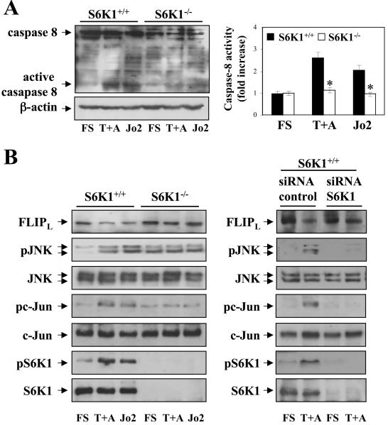
Figure 3. S6K1 deficiency protected neonatal hepatocytes from caspase-8 activation by inhibiting JNK-mediated FLIPL degradation.
S6K1+/+ and S6K1−/− immortalized hepatocytes were stimulated with TNFα (30 ng/ml) plus actinomycin D (100 ng/ml) (T+A) or Jo2 (2 μg/ml) for 16 h in DMEM plus 5% FS. A. (left panel) Total protein (50 μg) was used for western blot analysis with the corresponding antibodies against caspase-8 and anti-β̣-actin as a control for protein loading. A representative experiment is shown. (right panel) S6K1+/+ and S6K1−/− immortalized hepatocytes were treated as described above and caspase-8 enzymatic activity was measured. Results are expressed as fold increase of caspase-8 enzymatic activity compared to the S6K1+/+ condition, which was arbitrarily assigned a value of 1, and are expressed as means ± SE from three independent experiments. Statistical significance was determined by Student's t test comparing the S6K1−/− condition with respective values for wild-type controls. *p<0.05 was considered significant. B. (left panel) Apoptosis was induced as described in A. Total protein (50 μg) was used for western blot analysis with the corresponding antibodies against FLIP, phospho-JNK (Thr183/Tyr185), total JNK, phospho-c-Jun (Ser73), total c-Jun, phospho-S6K1 (Thr 389) and total S6K1. A representative experiment is shown. (right panel) S6K1+/+ immortalized hepatocytes were seeded in 6 cm dishes and incubated overnight at 37°C with 5% CO2. When 40- to 50% confluence was reached, cells were transfected with 100 nM of control or siRNA oligos following DharmaFECT General Transfection Protocol. After 48 h, hepatocytes were treated as described above. Then, cells were collected and cell lysates were analyzed by western blot with the indicated antibodies. Representative autoradiograms are shown.

