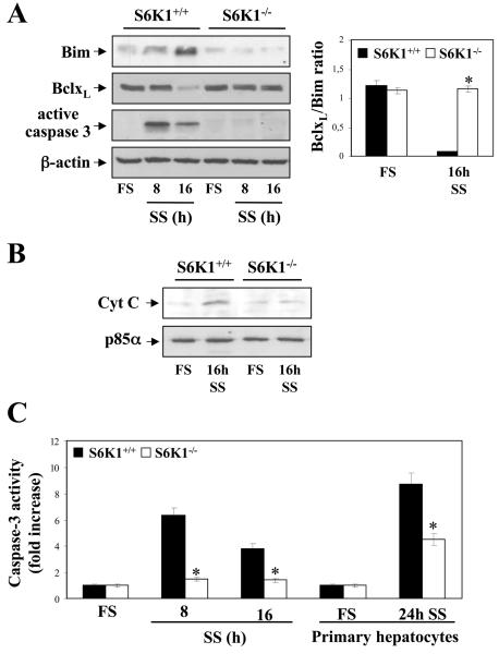
Figure 7. S6K1 deficiency protected neonatal hepatocytes from cytochrome C release and caspase-3 activation upon serum withdrawal.
S6K1+/+ and S6K1−/− immortalized hepatocytes were incubated in serum-free medium for 8 and 16 h. A. (left panel)Total protein (50 μg) was used for western blot analysis with the corresponding antibodies against Bim, BclxL and active caspase-3. The anti-β̣-actin antibody was used as a loading control. A representative experiment is shown. The autoradiograms corresponding to three independent experiments were quantitated by scanning densitometry and the BclxL/Bim ratio was calculated. Results are expressed in arbitrary units. Statistical significance was determined by Student's t test. *p<0.05 was considered significant (right panel). B. Mitochondria were separated from cytosol and cytochrome C content was analyzed in the cytosolic fraction by western blot. The anti-p85α antibody was used as a loading control. A representative experiment is shown. C. S6K1+/+ and S6K1−/− primary and immortalized hepatocytes were treated as described above and caspase-3 enzymatic activity was measured. Results are expressed as fold increase of enzymatic activity as compared to the S6K1+/+ condition, which was arbitrary assigned a value of 1, and are means ± SE from three independent experiments. Statistical significance was determined by Student's t test. *p<0.05 was considered significant.

