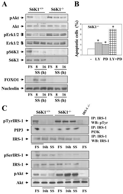
Figure 8. Survival pathways remain intact in S6K1-deficient hepatocytes upon growth factors withdrawal.
S6K1+/+ and S6K1−/− immortalized hepatocytes were incubated in serum-free medium for 8 and 16 h. A. At the end of the culture time, attached and non-attached cells were collected and total cell lysates and nuclear extracts were prepared. (upper panel) Total protein (50 μg) was used for western blot analysis with the corresponding antibodies against phospho-Akt (Ser 473), total Akt, phospho-ERK1/2 (Thr 202/Tyr 204), total ERK1/2, phospho-S6K1 (Thr 389) and total S6K1. (Lower panel) Nuclear protein (50 μg) was submitted to western blot analysis with the anti-FOXO1 antibody and the anti-nucleolin antibody as a loading control. A representative experiment is shown. B. S6K1-deficient hepatocytes were treated with LY 294002 (40 μM) and PD 098059 (20 μM), alone or in combination with serum-free medium for 16 h. The percentage of cells with DNA lower than 2C (apoptotic cells) was determined by flow cytometry as described in Materials and Methods. Statistical significance was performed by Student's t test comparing the values of S6K1−/− hepatocytes cultured in serum-free medium plus LY, PD or LY plus PD with the respective values of S6K1−/− cells cultured in serum-free medium. *p<0.05 was considered significant. C. Apotosis was induced as described above. (Upper panel) At the end of the culture time, cells were lysed and 600 μg of total protein were immunoprecipitated with anti-IRS-1 antibody, and analyzed by western blot with anti-pTyr or used for an in vitro PI 3-K assay. The conversion of PI to PIP3 in the presence of [γ32-P] ATP was analyzed by TLC (left panel). (Lowert panel) Total protein (50 μg) was used for western blot analysis with the corresponding antibodies anti-phospho-IRS-1 (Ser307), anti-total IRS-1, anti-phospho-Akt (Ser 473) and anti-Akt. The anti-β̣-actin antibody was used as a loading control (right panel). Representative autorradiograms are shown.

