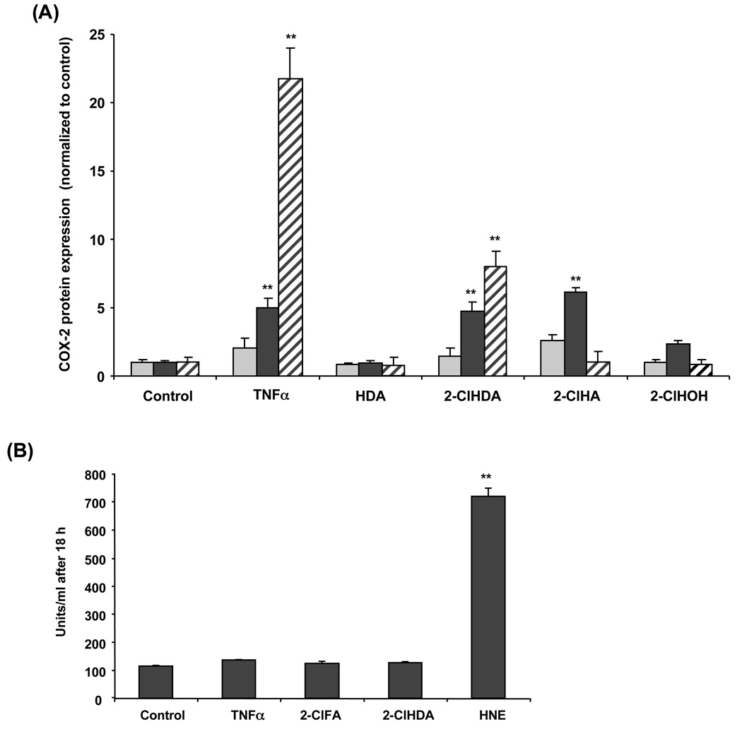
Figure 2. Time course of 2-chlorohexadecanal and 2-chlorohexadecanoic acid elicited COX-2 protein expression in HCAEC.
Cell lysates were prepared from HCAEC treated with ethanol vehicle, 50 ng/mL TNF-α, or 50µM of hexadecanal (HDA), 2-chlorohexadecanal (2-ClHDA), 2-chlorohexadecanoic acid (2-ClHA), or 2-chlorohexadecanol (2-ClHOH) for 2 h (light grey bars,  ), 8 h (black bars,
  ), and 20 h (striped bars,
  ). Samples were subjected to SDS-PAGE and western blotting for COX-2 expression and analyzed by densitometry of immunoreactive bands (A). Following COX-2 analysis, blots were subsequently stripped and then subjected to western blotting for β-actin, which was used to normalize each experimental analysis of COX-2 expression. Cell medium was assayed for general cell death using a lactate dehydrogenase release assay (B). Values (mean ± SEM) are expressed as fold increase over control-treated cells for three independent determinations, (**) represents p<0.01.

