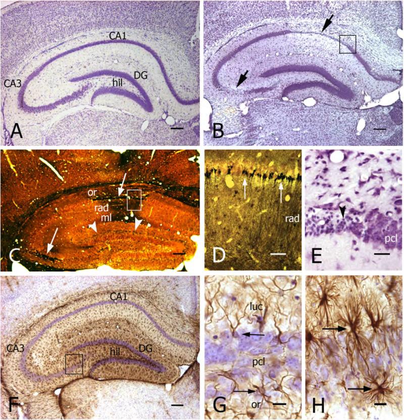
Figure 2. Chronic seizures in Kv1.1−/− mice are associated with severe pathohistological changes in hippocampus.
A, B) Transverse sections of the hippocampus from a 10 week-old wild-type mouse (A) and an age-matched Kv1.1−/− mouse (B). Cresyl violet staining shows the normal histological pattern of hippocampal regions and laminae in A. In the knock-out (B), there was significant neuronal cell loss in CA1 and CA3b/c subfields (arrows).
C) Neuronal cell death and degeneration (particularly in CA1 and CA3 - white arrows) are shown in a Fink-Heimer-stained transverse hippocampal section from a 10 week-old Kv1.1−/− mouse. Note also terminal staining in the dentate inner molecular layer (arrowheads), reflecting hilar mossy cell degeneration. Abbrevations: or, stratum oriens; rad, s.radiatum; ml, s.moleculare.
D) Higher magnification of the CA1 region (indicated area in C) showing degenerated CA1 pyramidal neurons within the pyramidal cell layer (black cell bodies) and terminal degeneration in strata radiatum (rad) and moleculare. Fink-Heimer staining.
E) Higher magnification of the indicated area in B, showing pyknotic pyramidal neurons (arrowhead) located in the CA1 pyramidal cell layer (pcl).
F) Transverse section of the hippocampus from a 5 month old Kv1.1−/− mouse, immunoreacted against GFAP. Note the high level of GFAP immunoreactivity (brown reaction product), particularly in the dentate gyrus (DG) and CA3c subfield. Cresyl violet counter-staining in F-H.
G, H) Photomicrographs comparing normal astrocytes (arrows in G) from CA3 of a wild-type mouse and reactive astrocytes (arrows in H, from boxed area in F) from a Kv1.1−/− mouse.
Scale bars: A-C, F, I, and J: 200μm; D and K: 50μm; E, G, and H: 20μm.

