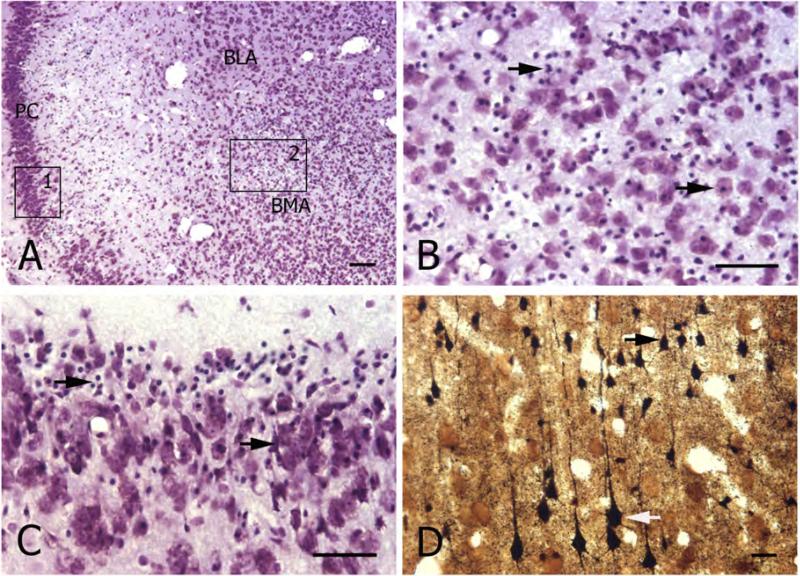
Figure 3. Chronic seizures in Kv1.1−/− mice are associated with pathological changes in multiple brain regions.
A) Transverse sections through the brain (level of the rostral hippocampus) of a 10 week-old Kv1.1−/− mouse who experienced status epilepticus. Cresyl violet staining shows severe cell loss in piriform cortex (PC) and anterior basomedial amygdaloid nucleus (BMA). For comparison, note the relatively preserved neuronal architecture in the anterior basolateral amygdaloid nucleus (BLA).
B) Higher magnification of the amygdala (BMA) (indicated area 2 in A) showing neuronal cell loss and many pyknotic cells (arrows).
C) Higher magnificationof the piriform cortex (indicated area 1 in A) showing severe neuronal damage, including pyknotic cells (arrows).
D) Higher magnification of a Fink-Heimer stained transverse section from the parietal neocortex (same animal as in A-C above) showing degenerated neurons in layer II/III (black arrow) and degenerated layerV pyramidal neurons (white arrow).
Scale bars: A and D: 100μm; B and C: 50μm.

