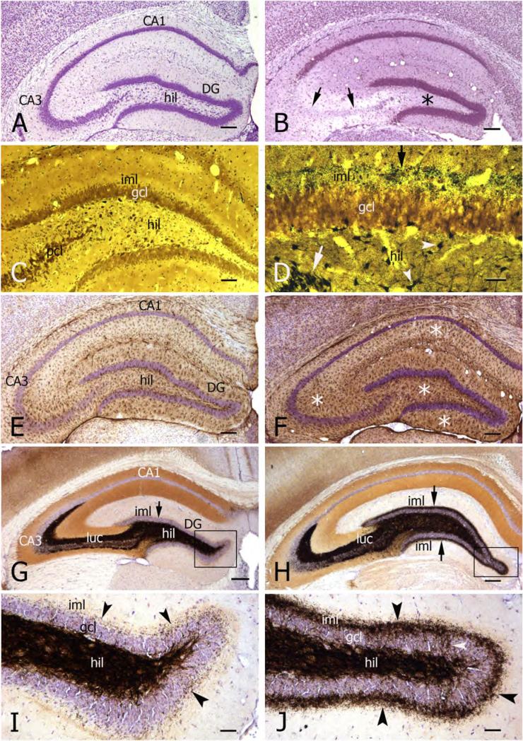
Figure 4. Variable pathologies in Kv1.1−/− mice reflect seizure severity.
A) Transverse section of the hippocampus (cresyl violet-stained) from a 9 month-old Kv1.1−/− mouse. This animal showed only mild seizure activity, and histology revealed no obvious morphological damage in hippocampal subfields.
Abbrevations: DG, dentate gyrus; hil, dentate hilus.
B) In comparison, a transverse section from a 10 week-old Kv1.1−/− mouse experiencing status epilepticus shows severe neuronal cell loss in CA3b and c subfields (arrows) and dentate hilus (asterisk).
C) Fink-Heimer staining of the dentate gyrus reveals little degeneration within the dentate hilus (black puncta) of a 10 month-old Kv1.1−/− mouse with mild seizure history. Abbrevations: iml, inner molecular layer; gcl, granule cell layer; pcl, CA3 pyramidal cell layer.
D) Fink-Heimer staining of a transverse section of the dentate gyrus of a 10 week-old Kv1.1−/− status epilepticus mouse reveals degenerated CA3 pyramidal cells (white arrow) and hilar neurons (white arrowheads, indicating degenerated mossy cells and interneurons), as well as severe terminal degeneration within the inner molecular layer (iml; black arrow).
E, F) Transverse sections of the hippocampus of a 10 month old (E) and a 10 week-old (F) Kv1.1−/− mouse. GFAP-immunostaining (cresyl violet counterstaining) shows moderate gliosis in dentate gyrus (DG) and hippocampal CA3 subfield in E (mild seizure history) compared to the pattern of severe gliosis in the dentate gyrus and all hippocampal subfields (asterisks) in F (status epilepticus history).
G) Transverse section of the hippocampus from a 9 month-old wild-type mouse showing the normal staining pattern of Timm-positive mossy fiber boutons, localized to dentate hilus (hil) and CA3 strata lucidum (luc) and oriens. Cresyl violet counterstaining in G-J.
H) Timm staining of a transverse section from a 5 month-old Kv1.1−/− mouse, showing mossy fiber sprouting into the dentate inner molecular layer (iml; arrows) as well as into the granule cell layer and s.oriens.
I) Higher magnification of the dentate crest (indicated area in G) from a wildtype mouse, showing sparse Timm-stained mossy fiber boutons in the granule cell layer (gcl) and inner molecular layer (iml; arrowheads). Abbreviation: hil, dentate hilus.
J) Higher magnification of the dentate crest (indicated area in H) from a Kv1.1−/− mouse, showing intense mossy fiber sprouting into the granule cell layer (gcl; white arrowhead) and inner molecular layer (iml; black arrowheads).
Scale bars: A, B, and E-H: 200μm; C: 100μm; D, I and J: 50μm.

