Figure 1.
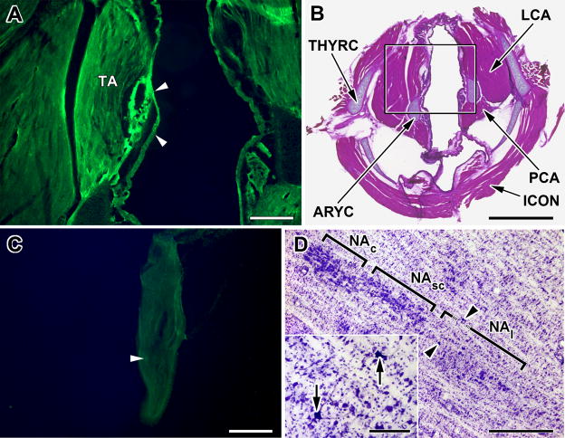
Pseudorabies inoculation in the thyroarytenoid (TA) muscle and transport to the nucleus ambiguus.
A. Fluorescent micrograph of a tranverse section through the rat larynx showing typical site of inoculation (arrowheads) of PRV-GFP into the thyroarytenoid muscle (TA) 60 hours after inoculation. Scale bar: 200μM.
B. Low power photomicrograph of the section shown in A now counterstained with hemotoxylin and eosin. The attachment of the thyroarytenoid muscle (TA) to the thyroid (THYRC) and arytenoid (ARYC) cartilages can be seen, as can the relative locations of the posterior (PCA) and lateral (LCA) cricoarytenoid and the inferior constrictor (ICON) muscles. Black square outlines the image shown in A. Scale bar: 1mm
C. Fluorescent micrograph of a longitudinal section through the nodose ganglion from an animal inoculated with PRV-GFP 60 hours prior to sacrifice. Note that only a single GFP fluourescent neurons (arrowhead) in this section. Scale Bar: 200μM
D. Cresyl violet counterstained horizontal section, 50 μM thick, through the rat medulla showing initial infection of motor neurons (arrowheads; arrows in inset) in the loose portion of the nucleus ambiguus (NAl) three days after inoculation of PRV into the TA. No infection was seen in the semi-compact (NAsc) or compact (NAc) parts of the nucleus ambiguus. Scale bar: 1mm; Scale bar in inset: 100μM.

