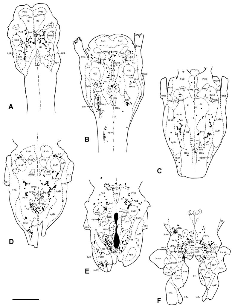
Figure 9.
Plots of infected neurons (black dots) in six representative 50μM thick horizontal sections through the pons and medulla from a sympathectomized animal sacrificed 5 days after inoculation of PRV into the TA. These plots show the typical early 5 day pattern. Section A is the most ventral and section F the most dorsal. Note the absence of infected neurons in the rostral ventrolateral medulla (RVL), A5 and area postrema (AP); these areas were almost always infected in non-sympathectomized animals. Scale bar: 2 mm

