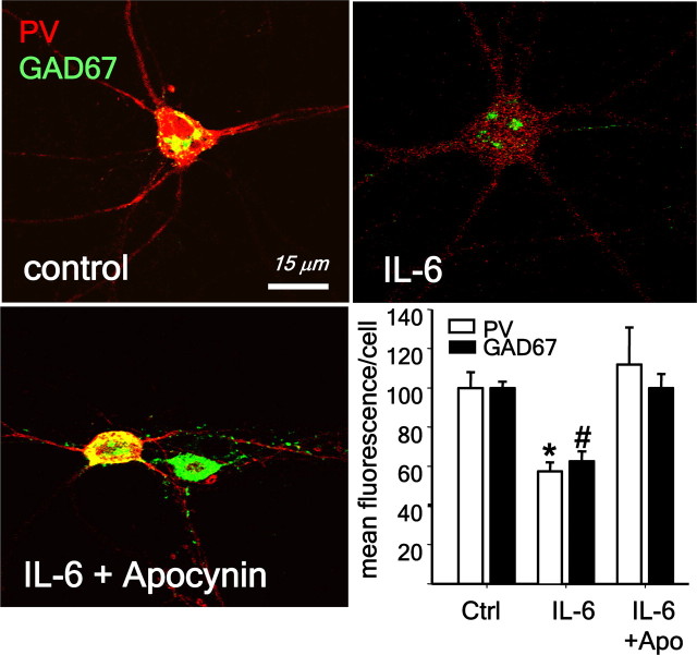
Figure 3.
IL-6 exposure leads to the loss of phenotype of PV-interneurons in primary neuronal cultures. Neuronal cultures were treated with IL-6 (10 ng/ml) in the absence (control) or presence of the Nox2 inhibitor apocynin (0.5 mm) for 24 h. Fluorescence confocal images of representative fields depicting the expression of PV and GAD67 in PV-interneurons. Bar graph represents the quantification of fluorescence expressed as percentage of control. (*p < 0.01, #p < 0.001. n = 4 experiments per condition.) Data are means ± SEM. Baseline intensities: PV = 135 ± 32; GAD67 = 114 ± 26.

