
Figure 3.

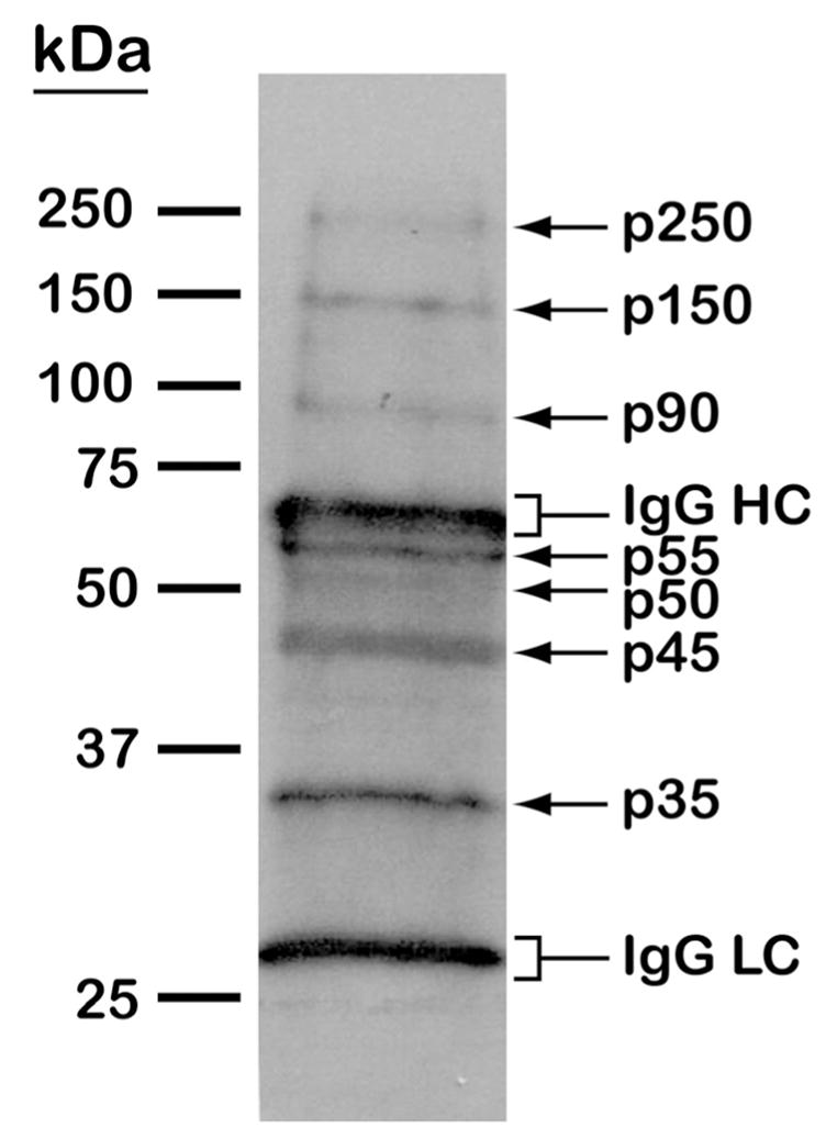
(A) MS/MS spectrum of the tyrosine-phosphorylated peptide LSPSEPNVAYpICSR from glycogen synthase kinase 3 (GSK3). Matched b and y fragments are indicated. (B) Western blotting analysis of tyrosine phosphorylated proteins in T. cruzi. Epimastigote extracts were subjected to anti-phosphotyrosine immunoprecipitation and separation by 10% SDS-PAGE. After blocking with BSA, the membrane was probed with the anti-phosphotyrosine antibody, followed by detection with horseradish peroxidase conjugated anti-mouse IgG and chemiluminescent reagent. Arrows denote T. cruzi tyrosine phosphorylated proteins. Brackets denote immunoglobulin G heavy (IgG HC) and light chains (IgG LC).