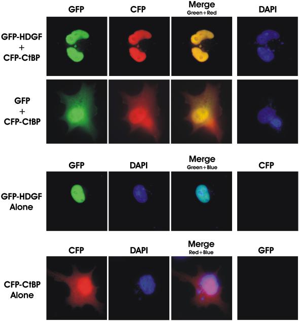
Figure 6.
HDGF induced the nuclear accumulation of CtBP. MCF-7 cells grown on cover slips were transfected with 0.5 ug of CFP-CtBP and GFP-HDGF (top panel) or GFP (2nd panel) for 48 hours, cells were fixed in 1% paraformaldehyde and the cover slips were mounted in DAPI containing medium, DAPI, CFP and GFP images are shown, with the merged images of CFP and GFP fluorescence (overlay). MCF-7 cells transfected with GFP-HDGF alone (3rd panel) or CFP-CtBP alone (bottom panel) was used as control. The merged image of GFP-HDGF (green) and DAPI (blue) in 3rd panel is showing the nuclear localization of GFP-HDGF. The image of the bottom panel is showing the diffused distributive pattern of CFP-CtBP in both nucleus and cytoplasm.

