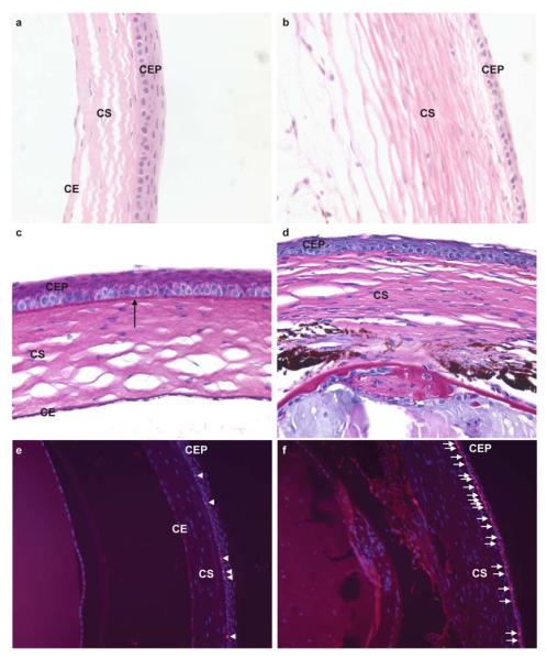
Figure 2. Le-Cre mediated deletion of Cited2 results in abnormal corneal morphology and increased corneal epithelial proliferation.

Histological examination was performed by the H&E staining of eye sections collected from 6.5-week old mice. Compared to the normal morphology of the Cited2flox/flox control cornea (a), thinner corneal epithelium and fewer corneal epithelial cells were observed in Cited2flox/flox;Le-Cre+ cornea (b). PAS staining was also performed in conjunction with H&E staining and revealed intact basement membrane in Cited2flox/flox control cornea (arrow in c), but not in Cited2flox/flox;Le-Cre+ cornea (d). Ki-67 immunostaining (red color) and DAPI counterstain (blue color) further showed increased Ki-67 positive cells (pink color merged from Ki-67 and DAPI staining) in Cited2flox/flox;Le-Cre+ corneal epithelium (arrows in f) compared to the few Ki-67 positive corneal basal cells detected in Cited2flox/flox control (arrowheads in e) (CEP: corneal epithelium; CS: corneal stroma; CE: corneal endothelium).
