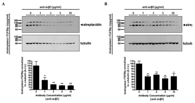
Figure 6. TGFBIp binds to HSFs by interacting with αvβ3 and αvβ5 integrins.

Western blot of cell lysates collected from HSFs preincubated with the function-blocking monoclonal antibodies (0 – 10 μg/ml) against αvβ3 (A) and αvβ5 (B) for 1 hr at 4 °C, before the addition of biotinylated rTGFBIp (10 μg/ml) for 5 hr at 4 °C. Blots were probed with streptavidin conjugated to alkaline phosphatase (upper panels), stripped and reprobed with α-tubulin antibody (lower panels). Arrowhead represents the biotinylated rTGFBIp (∼68 kD). Histograms represent the relative band intensities quantified, and each band was normalized to their corresponding α-tubulin. Data are expressed as the mean ± SEM by the Student's t-test for unmatched pairs for three individual experiments in triplicate (*p < 0.05, **p < 0.01).
