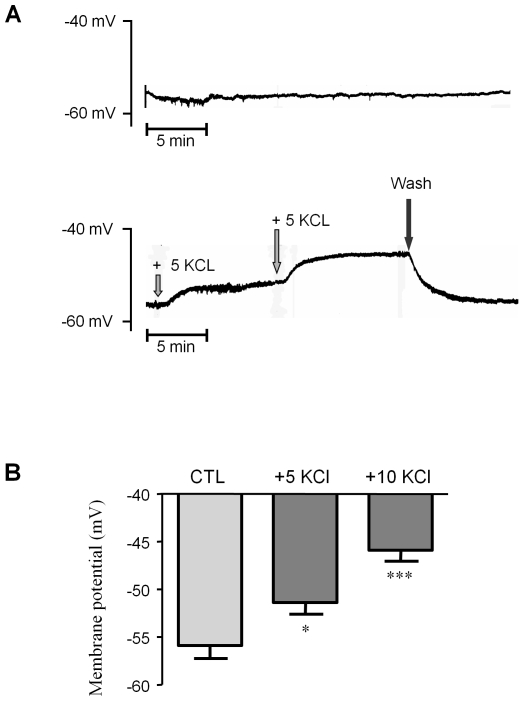
Figure 5. Membrane potential determination.
Membrane potentials were recorded in the SMCs of aortic rings under basal conditions and after addition of low KCl concentrations, using glass microelectrodes. (A) Representative recordings illustrating membrane potential stability in control experiments (upper panel) and depolarizations induced by the cumulative addition of 5 mmol/L KCl (lower panel). (B) Averaged membrane potentials under basal conditions (CTL) and after addition of 5 mmol/L and 10 mmol/L KCl (as described). *p<0.05; ***p<0.001; two-way ANOVA followed by a Bonferroni post-hoc analysis. Values represent means ±s.e.m. of 4 animals, with experiments performed in duplicate.

