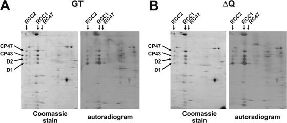
FIG. 3.
Comparative 2-D BN/SDS-PAGE analysis of pulse-labeled crude membrane extracts isolated from the Synechocystis sp. strain PCC 6803 GT and the quadruple mutant strain, ΔQ. Coomassie blue-stained gels and autoradiograms of the 2-D BN/SDS-PAGE analysis of pulse-labeled samples (an amount of 6 μg of Chl a was loaded per sample) were prepared as described by Sobotka et al. (43). The positions of PSII dimers (RCC2), PSII monomers (RCC1), and RC47 protein complexes (RC47, PSII core complexes lacking CP43) as well as those of the D1, D2, CP43, and CP47 PSII subunits are indicated.

